The Diaphragm

The diaphragm, viewed from above at left with the front of the body on top, is a sheet of muscle and tendon the divides that torso in two. Above is the thorax with the lungs and heart, below is the abdomen, featuring the intestines, stomach, liver, kidneys... It is dome shaped, slightly higher on the right side and it curves up toward the centre.
The diaphragm features a boomerang shaped central tendon which is connected all around by muscular fibers which originate on the lumbar spine,the bottom edge of the ribcase and sternum. The central tendon, or aponeurosis, is very strong. The diaphragm has three "holes", one each for the aorta, the esophagus and the vena cava. In the image above, the vena cava is blue, the aorta is red and the esophagus is yellow (in the middle).

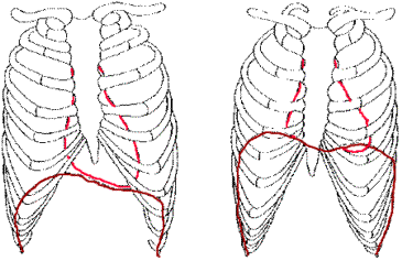
The image above features the ribs, sternum and costal cartilages in black
with the diaphragm in deep red and the heart in bright red. On the left, we
see the position of the heart and diaphragm at the peak of inhalation; on
the right we see the relaxed diaphragm just finishing the respiration cycle.
As you can see, the heart, which is attached to the diaphragm via its pericardium
(a membranous sac that envelops the heart), moves up and down with the diaphragm.
Also noticeable is the double dome effect of the diaphragm, where the diaphragm
is higher on the right side than the left, allowing the liver to be tucked
up under the bottom edge of the right ribcase, while the left is lower, allowing
more room for the heart.
Back to Respiration
- More on Anatomy
- The Physics of Breathing
- Application to the Performer

